Homework questions to be emailed to valenciabiologyhw@gmail.com
- Distinguish
between prokaryotic and eukaryotic cells
- Describe the
structure of a mitochondrion and explain the importance of
compartmentalization in mitochondrial function
- Distinguish among
amyloplast, chromoplast, and chloroplast
- Explain how
hydrophobic interactions determine membrane structure and function
- Explain how
active transport differs from diffusion
Notes on the Cell and Cell Membrane
• . XI. METHODS FOR STUDYING CELL STRUCTURE --
• How do we look at cells?
• A. Microscopy: whole tissues, cells, parts of cells
• 1. Light Microscopes -- use visible light
• Light microscopes (LMs)
– Pass visible light through a specimen
– Magnify cellular structures with lenses
• Electron microscopes (EMs)
– Focus a beam of electrons through a specimen (TEM) or onto its surface (SEM)
• The scanning electron microscope (SEM)
– Provides for detailed study of the surface of a specimen
• The transmission electron microscope (TEM)
– Provides for detailed study of the internal ultrastructure of cells
Some Important Dates
Some Important Dates
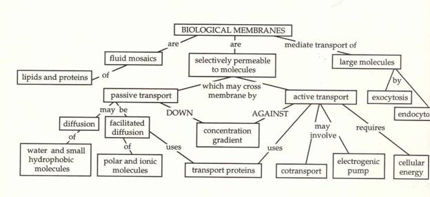
• . II. Membrane Functions
• A. Form selectively permeable barriers
B. Transport phenomena
• 1. Passive diffusion
• 2. Mediated transport
• a. facilitated diffusion
• (1) carrier proteins
(2) channel proteins
• (a) non-gated channels
(b) gated channels
• . b. active transport
• C. Cell communication and signaling
D. Cell-cell adhesion and cellular attachment
E. Cell identity and antigenicity
F. Conductivity
• . III. Fluid Mosaic Model of Membrane Structure
•
• . A. Mosaic: an object comprised of bits and pieces embedded in a supporting
structure
• 1. membrane lipids form the supporting structure
2. membrane proteins provide the bits and pieces
3. both lipids and proteins may be mobile or 'fluid'
• . B. Membrane lipids: the supporting structure
• 1. phospholipids
2. glycolipids
3. cholesterol
• C. Membrane proteins: the bits and pieces
• 1. integral (intrinsic) proteins
2. peripheral (extrinsic) proteins
• . IV. The Membrane Lipids
• A. phospholipids
• 1. most abundant of the lipids in membranes: form a lipid bilayer
2. phospholipid composition
• a. glycerol backbone covalently linked to:
b. two long, non-polar fatty acid hydrocarbon chains
c. variable phosphate-containing polar group
• 3. phospholipids are amphiphilic (amphipathic) molecules:
• a. hydrophobic ('water fearing') end: fatty acid chains
• orient toward the interior of the membrane
• b. hydrophilic ('water 'loving') end: phosphate group end
• orients towards the extracellular space or cytoplasm
• .
• 5. synthesis occurs in the membranes of the endoplasmic reticulum
• All of the phospholipids are initially synthesized on the cytoplasmic side of
the lipid bilayer. The phospholipids containing choline tend to get flipped to
the opposite face of the lipid bilayer (which is topologically equivalent to the
extracellular space) by enzymes known as 'flippases'. Once 'flipped', further
flip-flopping is rare.
• .
• B. Glycolipids
• 1. least common of the membrane lipids (~2%)
2. always found in outer leaflet of plasma membrane*
• 3. general structure of a glycolipid is a variation on the phospholipid
theme
• a. two long hydrocarbon chains
• (1) hydrophobic, non-polar part of molecule
• b. carbohydrate component: one or more sugars
• (2) hydrophilic, polar part of molecule
• 4. synthesis of glycolipids
• a. starts in membranes of endoplasmic reticulum
b. carbohydrates added in Golgi apparatus
• .
• C. Cholesterol
• 1. steroid; lipid soluble; found in both leaflets of lipid bilayer
2. amphiphilic: -OH group forms the polar end of the molecule
3. synthesized in membranes of endoplasmic reticulum
• .
• . V. Membrane Proteins
• A. Integral (intrinsic) proteins
• 1. penetrate the bilayer or span the membrane entirely
2. can only be removed from membranes by disrupting the phospholipid bilayer
3. types:
• . a. transmembrane proteins
• (1) single-pass
(2) multiple-pass
• Trans-membrane proteins have membrane spanning portions containing alpha
helically arranged sequences of 20-25 hydrophobic amino acids. Short strings of
hydrophilic amino acids separate the hydrophobic sequences from each other:
• . These hydrophilic stretches tend to be found exposed to the more aqueous
environments associated with the cytoplasm or the extracellular space
• . VI. Membrane Dynamics
• A. Lipid Asymmetry
• 1. phospholipids are asymmetrically distributed within the lipid bilayer
• a. outer leaflet of plasma membrane
• (1) phosphatidylcholine
(2) sphingomyelin
• . b. inner leaflet of plasma membrane
• (1) phosphatidylethanolamine
(2) phosphatidylserine*
• *carries a net negative charge
• 2. glycolipids exclusively found on outer half of membrane
• B. Lipid Mobility
• 1. mobility of membrane lipids:
• a. rotational movement
b. lateral movement
• .
• . The lipids (especially the phospholipids) are mobile within their half of
the lipid bilayer. Flip flop to the opposite side of the membrane is rare.
Mobility of the phospholipids tends to increase as the number of double bonds
('kinks') between adjacent carbon atoms in the fatty acid chains increase.
• 2. cholesterol effects on membrane fluidity:
• At high temperatures cholesterol tends to reduce membrane fluidity, probably
by interacting with the hydrocarbon tails of the phospholipid and glycolipid
molecules. At low temperatures cholesterol helps prevent membranes from freezing
and thus tends to maintain membrane fluidity
• .
• . 2. carbohydrates of glycoproteins always at outer surface
• a. help form the 'glycocalyx' (along with glycolipids
• . D. Protein Mobility
• 1. rotational mobility
2. lateral diffusion
• Protein mobility can vary greatly. Some proteins are free to move. Others may
be tethered to structures in the cytoplasm or extracellular spaces, thus
restricting their movement. Some types of cell junctions (e.g., tight junctions)
can restrict protein movements to a specific membrane domain.
• E. Membrane Protein Conformational Changes
• 1. help explain many many membrane functions
2. concept:
• The binding of a membrane protein/glycoprotein to some other cellular or
extracellular substance, molecule, ion, etc. can/will result in a 3-dimensional
conformational change in that membrane protein.
• . That conformational change can/will, in turn, drive or inhibit some other
cellular event.
• .
• Example 1: facilitated diffusion
• Example 2: signaling
•
• .. Membrane lipids: the supporting structure
• 1. phospholipids
2. glycolipids
3. cholesterol
• C. Membrane proteins: the bits and pieces
• 1. integral (intrinsic) proteins
2. peripheral (extrinsic) proteins
• . b. active transport
• C. Cell communication and signaling
D. Cell-cell adhesion and cellular attachment
E. Cell identity and antigenicity
F. Conductivity
• http://www.d.umn.edu/~sdowning/Membranes/lecturenotes.html
• . C. Protein Asymmetry
• 1. many different kinds of proteins are in the cell membranes
• a. each type has a unique conformation and orientation
b. flip flop of proteins does not occur
c. conformational changes of protein can occur
• b. covalently tethered integral membrane proteins
• Tethered integral membrane proteins may be largely exposed to either the
cytoplasm or aqueous extracellular space, but are covalently linked to membrane
phospholipids or glycolipids
• .
• . . many integral proteins are glycoproteins
• a. covalently linked via asparagine, serine, or threonine to sugars
• The sugars of glycoproteins are exclusively found on the extracellular side of
the membrane or topological equivalent
• . 5. synthesis of integral proteins:
• a. occurs in the rough endoplasmic reticulum
b. many integral proteins wind up as glycoproteins
• (1) glycosylation begins in lumen of er
(2) carbohydrates are modified in Golgi
• . 6. integral proteins often form protein complexes having multiple subunits
• 7. functions
• a. enzymatic
b. receptors
c. transport
d. communication
e. adhesion
• B. Peripheral (extrinsic) proteins
• 1. do not penetrate the phospholipid bilayer
2. are not covalently linked to other membrane components
3. form ionic links to membrane structures
• a. can be dissociated from membranes
b. dissociation does not disrupt membrane integrity.
• 4. located on both extracellular and intracellular sides of the membrane
• a. often link membrane to non-membrane structures
• 5. synthesis of peripheral proteins:
• a. cytoplasmic (inner) side: made in cytoplasm
b. extracellular (outer) side: made in er and exocytosed
• .
• . I. Cell types
• A. Prokaryotes-no nucleus and (generally) no internal membranes
• B. Eukaryotes-nucleus and organelles bounded by internal membranes
• II. Eukaryotic Cell Structure
• I. Cell types
• A. Prokaryotes-no nucleus and (generally) no internal membranes
• B. Eukaryotes-nucleus and organelles bounded by internal membranes
• II. Eukaryotic Cell Structure
• A. Prokaryotes: Simplest organisms; unicellular. Bacteria, Cyanobacteria (bluegreen
algae ---> photosynthetic), and Archaebacteria ... 1. No internal membranes -->
no nucleus
•
• a) plasma membrane surrounded by cell wall; plasma membrane segregates cell
from its environment but is selectively permeable allowing some molecules to
cross
• b) DNA coiled into region called nucleoid
• 2. External Structures
•
• a) pili --> cell-cell attachment and communication
• b) flagella --> cell movement
• . B. Eukaryotes: larger and more complicated; both unicellular and
multicellular
• . C. Size ... 1. Procaryotes are small: a) mycoplasma -- 0.1 - 1 µ b) bacteria
-- 1 - 10 µ
•
• 2. Eukaryotes are larger: 10 - 100 µ
•
• 3. What limits cell size -- Surface/Volume
• . a) must take up nutrients and O2 by diffusion across plasma membrane
• b) cell volume increases as (cell dia)3 ==> as cells become too large their
plasma membrane surface area becomes inadequate to supply cell needs.
• . 4. Eukaryotes take some functions of the plasma membrane of bacteria and
segrate them in internal membranes of organelles.
Eukaryotic Cell Strucutre
• . A. Endomembrane system
• 1. Nucleus
• nuclear membrane: --double membrane lined by nuclear lamina and nuclear pores
• DNA + special proteinË chromosomes
• Nucleolus-where ribosomes are made
• . XIII. ENDOMEMBRANE SYSTEM: ...
• internal membranes of eukaryotic cells related by direct physical contact or
by transfer of membrane segments via small vesicles; derived from Endoplasmic
Reticulum.
• . A. Nucleus ...
• 1. Nuclear Envelope
•
• a) double membrane surrounding the nucleus separating it from the rest of the
cell
• b) nuclear pores - pores through both membranes; formed by protein molecules;
controls traffic of molecules between the cytoplasm and the nucleus
• c) nuclear lamina - layer of proteins inside the nuclear membrane; may be
responsible for stabilizing the nuclear membrane.
• d) Nucleolus--where ribosomes are made
• 2. Chromosomes
• a) DNA + proteins (histones) ==> chromatin which is dispersed through most of
the nucleus. The proteins keep the very long DNA molecules organized. Chromatin
is divided into individual units called chromosomes.
• b) during cell division chromatin segregates into individual chromosomes
• c) number of chromosomes depends upon type of cell; e.g. what organism and
whether it is somatic or germ (reproductive) form. .
• . 3. Nucleolus -- part of the nucleus responsible for synthesis of ribosomes
needed for protein synthesis in the cytoplasm. Nucleolar Organizers are regions
of chromosomes which contain multiple copies of genes for ribosome synthesis
Endoplasmic Reticulum
• B. Endoplasmic Reticulum:
• half of the total membrane in many cells--produces membrane for others
• 1. Lumen -- space enclosed by ER; separate from the cytoplasm; contains
proteins targeted for
• a) secretion; b) other organelles
• 2. ER is continuous with the Nuclear Envelope -- nuclear memb. is part of the
endomembrane syst.
• .
• 3. Rough ER -- contains many bound ribosomes which bind to the ER after
beginning synthesis of proteins which are to be secreted into the lumen. These
proteins are identified by a short signal sequence at their N-terminus which
causes the ribosomes to bind to the ER. The proteins are then threaded through
the ER membrane as they are synthesized.
• 4. Smooth ER -- connected to rough ER; no ribosomes
• .
• . a) formation of vesicles to transport membrane and contents of lumen;
vesicles bud off ---> Golgi
• b) modified in special cells for various functions - e.g. sarcoplasmic
reticulum in muscle cells.
• c) lipid synthesis
• d) drug detoxification
• 2. Endoplasmic Reticulum: --double membrane continuous with nuclear
membraneËER lumen separated from cytosol
• Rough ER contains bound ribosomes
• Smooth ER no ribosomes
• A. Ribosomes -- for synthesizing proteins
• 1. Large assemblies of RNA + dozens of different proteins, synthesized in
nucleolus in eukaryotes
•
• ribosomal subunits - 2 (large and small) form an intact unit only when
synthesizing protein
• eukaryotic - 20 nm x 30 nm; prokaryotic ribosomes are slightly smaller
• . 2. Location:
•
• found free in the cytoplasm in both eukaryotes and prokaryotes
• bound to endoplasmic reticulum in eukaryotes ==> rough ER
• polysomes - in both cases several - many ribosomes are frequently found
together bound to the same RNA molecule all synthesizing the same protein. .
Golgi Appratus
• 3. Golgi Apparatus
• communicates with ER via transport vesicles that bring membrane and proteins
synthesized in ER
• Golgi modifies lipids and proteins and sends them on
• 4. Lysosomes - digest macromolecules using enzymes transported from Golgia via
transport vesicles
• C. Golgi Complex ...
• 1. Intermediate step for vesicles produced by smooth ER transporting membrane
and proteins
• a) flattened discs of stacked membranes enclosing a lumen
• b) cis face - end of Golgi where vesicles arrive; very close to or in contact
with ER
• c) trans face - opposite end from forming face; where processed vesicles are
leaving
• 2. Golgi modifies products of ER
• .
Other Structures
• D. Lysosomes -- Made from ER ---> Golgi ---> Lysosomes ...
• 1. Compartment surrounded by membrane which contains enzymes to digest
(hydrolyze) macromolecules, proteins polysaccharides, fats, nucleic acids. .
• a) membrane keeps enzymes from digesting functional parts of the cell
• b) maintains low pH (approx. 5) where these digestive enzymes are most active
• 2. Used to digest:
•
• a) external material by engulfing into vesicles (Phagocytosis) which then fuse
with lysosomes.
• b) digest and recycle cellular materials -- engulfs damaged organelles
• . Peroxisomes: Not part of Endomembrane System and not Energy Transducing
• 1. fatty acid degradation
• 2. H2O2 converted to H2O + O2 by an enzyme, catalase
• E. Vacuoles: large vesicles used to segregate some compounds from the rest of
the cell
• 1. Food Vacuoles -- produced by phagocytosis (plasma membrane enveloping large
particles)
• 2. Plant Central vacuoles ... a) storage of organic compounds (including
proteins) and inorganic ions
• b) serves the functions of lysosomes in plant cells
• .
• 5. Vacuoles; commonly found in plant cells and yeast taking the place of
lysosomes
• 6. Plasma Membrane
• receives protein and lipid synthesized in ER and modified in Golgi
communicates via transport vesicles
• .
Other Structures
• ENERGY TRANSDUCING ORGANELLES
• A. These are different from the Endomembrane System.
• They have evolved from prokaryotes engulfed by primitive eukaryotes ==>
symbiotic relationship where each benefits
• .
• . B. Energy Transducing Organelles--evolved from prokaryotes and have their
own DNA, ribosomes etc.
• 1. Mitochondria
• 2 membranes and two compartments
• oxidize food molecules to produce ATP
• 2. Chloroplasts
• 3 membranes and 3 compartments
• adsorb light and use energy to produce carbohydrate from CO2
• 1. component proteins are synthesized on free cytoplasmic ribosomes
•
• 2. Have their own genetic system, DNA and ribosomes
•
• 3. They grow and divide independently of the other organelles
• .
Mitochondria
• . B. Mitochondria ... 1. Cellular Respiration -- oxidize intermediate products
of metabolism to CO2 and H2O; excess free energy transformed into synthesis of
ATP
•
• 2. Structure
• a) Approximately 1 µ in diameter; may be very long in some cells
• b) 2 membranes - inner membrane and outer membrane
• c) inner membrane has large surface area and folds inward forming cristae;
contains membrane proteins responsible for respiration and ATP synthesis
• d) outer membrane is a sieve permeable to small molecules; intermembrane space
is similar to cytoplasm in concentration of small molecules
• e) matrix - contains enzymes responsible for many steps of metabolism, DNA,
ribosomes, etc.
• .
• C. Chloroplasts (member of a class of plant organelles called plastids) 1.
Photosynthesis
•
• a) light energy used to make ATP via mechanism similar to that in
miotochodnria
• b) ATP is used to make CH2O from CO2 and H2O
• .
Chloroplast
• . 2. Structure
• a) 2 - 5 µ in diameter
• b) 3 membranes - outer membrane, inner membrane, and thylakoid membrane
• c) stroma - within inner membrane; contains enzymes for metabolism, DNA,
ribosomes, etc.
• d) thylakoids - disc-like sacks which stack producing grana. Contain membrane
proteins which absorb light and make ATP.
• D. Cytoskeleton--maintains cell shape and gives cells motility
• 1. Microfilaments: --thinnest; polymers of globular protein actin; can
assemble and disassemble (a.k.a. Actin Filaments)
• component of muscle
• used in cell movement and cell division
• .
• . CYTOSKELETON: 3 different types of fibers ... A. Functions
• 1. Mechanical Support -- maintain cell shape
•
• 2. Motility
• a) movement of entire cell via pseudopods or flagella
• b) movement of organelles within cells
• c) separation of chromosomes
• d) cytokinesis -- division of cytoplasm
• (c) and (d) are essential for cell division
• .
• B. Microtubules:
• 1. Hollow rods approx. 25 nm in diameter
•
• a) constructed from globular proteins.
• b) Grow, usually, from addition of subunits to one end.
• .
• 2. Microtubules: --largest, cylinders of globular tubulin dimers; can assemble
and disassemble
• component of cilia and flagella
• used in separated chromosomes during cell division
• .
• 2. MT's cause:
•
• a) separation of chromosomes during Mitosis (cell division)
• b) movement of organelles and vesicles
• c) movement of cells -- cilia and flagella
• .
• 3. Centrioles -- bundles of 9 triplets of MT's at center of MT organizing
centers; these act as the focal point for MT formation ==> MT's radiate from
these organizing centers and separate the chromosomes during mitosis. Centrioles
are not essential; plants don't have them and mitosis still occurs.
• .
• . 4. Cilia and Flagella --
• a) central pair of MT's surrounded by 9 double MT's
• b) bend by action of dynein, and enzyme which gets energy from ATP; causes
cellular motion or motion of fluid past tissues
• c) attached to cell via basal body which is identical in structure centriole
• C. Microfilaments: Actin (globular protein which makes thin filaments in
muscle).
• 1. 7 nm diameter -- helical fiber made of globular actin monomers
•
• 2. Movement caused by another protein, myosin (makes thick filaments in
muscle)
•
• 3. Found in all types of eukaryotic cells and is responsible for
•
• a) constriction of plasma membrane during cell division -- cytokinesis
• b) pseudopod extension in ameboid movement
• c) contraction of muscles
• 3. Intermediate filaments: --intermediate in size; composed of fibrous
proteins rather than globular; generally does not disassemble once formed
• .
• . E. Motor Molecules--move along filaments using energy from ATP hydrolysis
• 1. Myosin--moves along Microfilaments (Actin Filaments)
• causes muscles to contract
• moves vesicles along microfilaments
• B. Motor Molecules ...Figure 4.25
• 1. One end attaches to "Cargo" which the motor molecule carries along "Rails"
of either Microtubules or Microfilaments
•
• 2. Fuel which powers the movement is a chemical called ATP which is produced
by cellular respiration.
• 2. Kinesin and Dynein: two different classes of molecules that move along
Microtubules
• dynein--causes movement of microtubules in cilia and flagella
• kinesin--causes movement of vesicles along microtubules
• F. Extracellular Matrix--external network of fibrous proteins connected to the
cytoskeleton through membrane proteins called Integrins .
• . CELL SURFACE INTERACTIONS
• A. Extracellular Matrix
• 1. Integrin: A membrane protein of the plasma membrane connects the
cytoskeleton with ...
•
• 2. Collagen and Fibronectin: extracellular glycoproteins
•
• B. Intercellular Junctions ...Figure 5.6
• 1. Tight Junctions: Join neighboring cells and prevent diffusion between them
•
• 2. Gap Junctions: Join the membranes of neighboring cells and allow diffusion
of small molecules from one cell to the other ==> Communicating Junctions
• G. Intercellular Junctions
• Tight Junctions--join neighboring cells
• Gap junctions--join cells and allow them to communicate
•
.