Cells 5/08
OBJECTIVES
The student should be able to:
|
1.Describe a typical plant cell. 2. Describe a typical animal cell and be able to differentiate it from a plant cell. 3. Describe the three main shapes of bacterial cells. 4. Differentiate a prokaryotic cell from a eukaryotic cell. |
||||
|
KEY TERMS |
|
|
|
|
|
a. animal cell |
j. |
cyanobacteria |
r. |
nucleolus |
|
b. bacteria |
k. |
diatom |
s. |
prokaryote |
|
c. bacillus |
1. |
eukaryote |
t. |
pseudopodia |
|
d. cell membrane |
m. |
flagellum |
u. |
protozoa |
|
e. cell wall |
n. |
green algae |
v. |
spirilla |
|
f. chloroplast |
o. |
hyphae |
w. |
sporangium |
|
g. cilia |
p. |
mycelium |
x. |
vacuole |
|
h. coccus i. colony |
q. |
nucleus |
y. zygospore |
|
Cells are thought to be the smallest, most basic unit of life in the same way that an atom is the smallest unit of an element. In today's laboratory we will examine cells of each of the five main kingdoms: Monera (bacteria), Protista (green algae and protozoa), Fungi (mushrooms and molds), Plants (Elodea), and Animals (buccal cavity smear). Please note that while the authors have used the mostly widely accepted taxonomic divisions, there are other methods of classification which result in six or eight kingdoms.
The cells that we will examine today will be studied with the use of the microscope, but all cells are not microscopic. The yolk of the egg is an individual cell and it can certainly be studied without the microscope. During your work today, you will learn to distinguish each type of cell from other related types. Most cells share many organelles, but in each cell type there are major differences of form and function. In this laboratory we will begin to examine the structure of cells and to compare and contrast cell types.
PROKARYOTIC CELLS
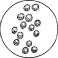
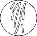
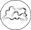
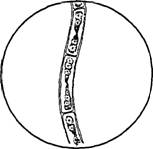
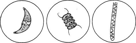
Prokaryotes are bacteria and blue-green algae. Prokaryotic cells are very small (.5-5 um) and are much less complex than eukaryotic cells. They are found as unicellular organisms, although they may appear in colonies, in bunches, or strings of cells. Monerans that we will look at today include various forms: cocci, bacilli, and spirilla. Some of these may be presented on prepared slides. Note that observed cell structure is not discernible with the techniques that you will be using today. Cell walls and capsules will be seen with stains. Remember that bacteria are very simple life forms but are ubiquitous and extremely important. Bacteria play a major role in decomposition of organic material, in disease as well as antibiotic production.
The bacteria that you will examine is found in yogurt with live cultures of bacteria. The bacteria commonly used are Streptococcus thermophilus, which ferments the sugar lactose, and Lactobacillus bulgaricus, which produces the flavors and aroma of yogurt.
COCCI BACILLI SPIRILLA
Draw the observed bacterial types.
PREPARE A SLIDE OF YOGURT CULTURE
|
1. |
Obtain a slide and cover slip. With a bacterial loop or a toothpick transfer a small amount of |
|
|
yogurt to the center of the slide. |
|
2. |
Smear the yogurt in an area slightly smaller than the cover slip. Allow it to dry. |
|
|
|
|
3. |
Place two drops of crystal violet or carbolfuchsin stain on the air-dried yogurt smear. |
|
4. |
Place a cover slip on the stained smear and examine under the microscope. |
 Draw the
bacteria you see in the yogurt
Draw the
bacteria you see in the yogurt
Cyanobacteria are blue-green algae. Blue-green algae is found in aquatic environments as well as in damp, terrestrial environments. They exist largely as colonies and filaments and produce spores that resist desiccation. They generally have gelatinous capsules that are often toxic and are not a normal food source for heterotrophs.
You may be using prepared slides or using fresh cultures of these organisms. A few cyanobacteria are Anabeana, Gloeocapsa, Oscillatoria, and Merismopedia. Your instructor may provide a survey culture of various organisms to key out and to examine.

ANABANA GLOEOCAPSA MERISMOPEDIA OSCILLATORIA
Draw and label several representatives of cyanobacteria.
EUCARYOTES
Eucaryotes include protists, fungi, plants, and animals. The characteristic that sets them apart from Monerans is the presence of a nucleus. Many other organelles are often present, such as mitochondria (cell respiration) and chloroplasts (photosynthesis in plant cells). The cells tend to be larger by at least a factor of 10 when compared to bacteria. Compartmentalization of venous cellular processes characterizes eucaryotes.
PROTISTA
Protista includes photosynthetic members, algae, heterotrophic single-celled organisms called protozoans, as well as some fungi-like protists. Protists are a food source for many organisms and comprise the base of the food chain. To examine algae we will use the green survey mixture in which a number of algae will be included. Note that algae cells have a cell wall made of cellulose. The plasma membrane is contiguous with the wall and will not be observed. These cells will also probably have visible chloroplasts, some of which will be oval, round, or spiral shaped. Much of the algae is either filamentous or colonial. While diagrams of a number of cell types with organelles are included in this lab, often the organelles will not be readily observed. Several drawings of algae have been included. (Note: Algae survey mixtures often contain cyanobacteria which are classified as Monerans.)
OEDOGONIUM CLOSTERIUM SCENEDESMUS SPIROGYRA
Key out and draw at least four algae representatives. Calculate cell size of each alga.
Protozoans differ in morphology considerably. We have chosen several representative species for you to examine: amoeba, paramecium, euglena. Note that each of these move differently. Amoeba extend cytoplasmic extensions in response to environmental stimuli. They use these pseudopodia (false-feet), extensions of cytoplasm, to move and to engulf prey. Paramecium use small oar-like structures called cilia, and euglena use flagella (longer cilia-like structures) to move through water. All of these microorganisms have a cell membrane. Note the drawings of these three and then record your own observations of the organism, calculating cell size as you do so.
Students record observations, label drawings, and calculate cell size.
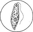
AMOEBA PARAMECIUM EUGLENA
FUNGI
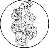
Fungi are heterotrophic multicellular organisms. Fungi include mushrooms, mildews, yeasts, and molds. Fungi are characterized by a vegetative structure called hyphae, which occur in mats called mycelia. Hyphae are filaments of cells. Fungi are ubiquitous and often grow in damp, dark places. A typical fungus is Rhizopus, which is a black terrestrial mold often found on old bread. There are sexual and asexual stages, which we will be examining today.
Examine a petri dish containing an asexually reproducing mycelium of Rhizopus using a dissecting scope. Locate the asexual spores-small black spheres in the sporangia that grow on erect sporangiophores.
Using a prepared slide of Rhizopus, examine the sexual stages. When hyphae of opposite mating types meet and fuse, a dark zygospore will be produced.
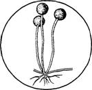
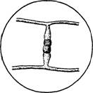
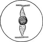
SPORANGIAPHORE GAMETANGIA ZYGOSPORE
The student should draw and label both asexual and sexual stages of Rhizopus.
Sporangiaphore Zygospore
KINGDOM PLANTAE
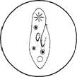
This kingdom includes mosses, ferns, horsetails, club mosses, conifers, angiosperms (flowering plants). To examine cell structure of a typical plant we will use Elodea, an aquatic plant. Place a leaf of Elodea on a slide. Use one drop of water and place the cover slip on the drop. Examine the visible structures and draw in the space provided. Locate the cell wall (which consists of cellulose), the chloroplasts, and the nucleus. Other organelles may not be visible.
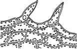
ELODEA
Draw and label the visible structures of Elodea.
With a razor blade, slice a very thin section of potato and stain with two drops of Logol's Solution. Look for leucoplasts, which are specialized starch-rich plastids. Draw a cell.

KINGDOM ANIMALIA
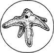
The kingdom animalia includes a variety of organisms such as nematodes, mollusks (clams), insects, fish, birds, and mammals. These are heterotrophic multicellular organisms with wide ranges of form and function.
We will examine a typical animal cell, a scraping from your cheek. With a toothpick scrape a small sample from the inside of your cheek in the oral cavity. Place a drop of methylene blue on the tissue and place a cover slip on the slide.
EPITHELIUM CELLS
The cells that you are examining will be pale blue with a nucleus that is dark blue. After examining the slide under low power, move to high power. Locate the plasma membrane, the nucleus, and the nucleolus, characterized by a darkened area in the nucleus. Calculate cell size and draw a few representative cells.
QUESTIONS
1. Differentiate between prokaryotic and eukaryotic cells.
2. Compare and contrast plant and animal cells.
3. Discuss methods of locomotion in Protozoans.
4. Describe the different types of Moneran cells.
5. Differentiate between sporangiophore and zygospore in Rhizopus