Phloem Lab
Staining Procedure
It is sufficient to stain the sections with
Aniline Blue.
Wait 5 min.
Rinse with water and observe.
Detailed protocol for Aniline Blue Staining
Place sections in IKI for 3 minutes,
Rinse with water
Stain 5 minutes with 0.1% aqueous aniline
blue.
Wash briefly with IKI
Mount in water.
|
The detailed
structure of sieve elements in the phloem cannot be observed easily without
the use of special staining techniques. Consequently, some of the material
used in this exercise will be fresh. Sections of living material are usually more
difficult to interpret than commercial slides. Therefore certain prepared
slides will be used for orientation, and to demonstrate the arrangement of
cells in the phloem, as well as the associations of phloem & xylem. |
|
|
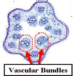
Primary Phloem of
squash (Cucurbita) Study prepared
slides of cross and longitudinal sections of Cucurbita
stems. Locate the xylem
and phloem. Does the phloem occur on one side of the xylem (collateral
bundle) or on both sides (bicollateral)???? Study hand
sections and stain with Toluidine Blue. Compare
these with the commercial slide. |
|
|
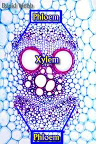
Cucumber
Vascular Bundle: It has Phloem on two sides of the Xylem Cucumber Vascular Bundle showing the Phloem.
The dark cells are Companion Cells & the largest cells are Sieve Tube
Members. |
|
|
High-power study
shows the three components of phloem tissue: Sieve Elements (here Sieve Tube
Members), Companion Cells (small cells accompanying the sieve elements), and Phloem
Parenchyma cells (intermediate in size between sieve elements and companion
cells). |
|
|
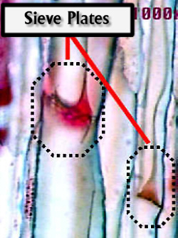
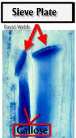
The end walls of
the sieve elements seen from the surface in cross sections,
bear highly differentiated Sieve Areas. These end walls are called Sieve
Plates. The protoplasts of adjacent sieve tube members form a continuum
through the sieve plates. |
|
|
These
connections are the Connecting Strands. Each is encased in Callose, a carbohydrate wall substance chemically
distinct from the cellulose that lines the sieve pores. |
|
|
Staining shows
that sieve elements appear end to end in longitudinal series and thus, form
Sieve Tubes. |
|
|
The lateral
walls of sieve tubes bear relatively undifferentiated Sieve Areas. The pores
in the sieve areas are much larger than typical pits and resemble those in
the sieve plate. |
|
|
Top view of a
Sieve Plate from a commercial slide at high magnification Commercial slide
of Phloem seen in Longitudinal section: Note the Red-Stained material which
contains Callose. also note the Sieve Plates |
|
|
In order to
demonstrate Callose in fresh material we will use
free-hand longitudinal sections of Cucurbita
stained with Aniline Blue. Aniline Blue preferentially stains callose. Furthermore, stained callose
emits fluorescence under ultra-violet and violet light. |
|
|
Callose will stain blue. However, Aniline Blue will also
stain other materials in the section so you need to locate the xylem which is
auto-fluorescent, then the phloem. Look for
concentrations of the stain in the phloem region, and locate the presence of
sieve plates in the highly stained areas. Callose accumulates
at the Sieve Plates due to the pressure that exists in the Phloem. |
|
|
Observe these
sections with a fluorescence microscope that clearly shows the sieve plates
because of aniline blue fluorescence. These will appear white or light blue
against a dark background. Plastids will
fluoresce red. Xylem fluorescence will also be blue but you can easily
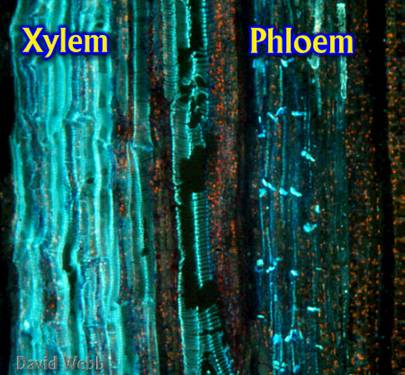
identify it due to the characteristic secondary wall thickenings. Overall view of
a longitudinal section of cucumber stem stained with Aniline Blue and seen with
Violet Fluorescence. The cell walls of the Xylem (left) are auto-fluorescent
while the fluorescence of the Phloem (right) is due to Callose
which has stained with Aniline Blue. |
|


|
The sieve plates
will be the most fluorescent areas because callose
accumulates there normally and becomes more concentrated after wounding. The
sieve plates vary in their orientation. Some are perpendicular to the long
axis of the stem while others may have 45O angles of inclination. The latter
can be seen in face view in longitudinal sections. This allows you to see the
sieve pores. Cucumber Phloem
stained with Aniline Blue & Viewed with Violet Light Fluorescence. |
|
|
Longitudinal
sections of Cucumis stems were stained with Aniline
Blue and photographed with a fluorescence microscope after excitation with
Violet Light. Callose in the Phloem is fluorescent and appears
bright under these conditions. It has a yellow/green color. This is partly
due to the blue color of the dye. The Sieve Plates
are especially fluorescent because they contain a lot of associated Callose and Callose accumulates
there upon injury. Lateral Sieve
Pores are also fluorescent. Xylem is autofluorescent and appears similar to the Phloem. Xylem
can be identified because of its characteristic secondary walls. Plastids appear
a red dots due to the fluorescence of Chlorophyll |
|


What else to do:
- Make two drawings only on
- Label your drawings based on what you
have learnt about the phloem.
•
Root
–
Zea, x.s. Slide
#100. Locate phloem, then draw sieve tube elements
and companion cells.
•
Leaf
–
Zea, x.s. Slide
#86. Note the pits on the vessel member walls
•
Stem
–
Cucurbita, x.s. & l.s. Material Box # 10.
•
First examine x.s. slides, figure out where phloem should be, and then
examine l.s. slides. Note
the sieve tube members and sieve plates.