Stems
Materials
1.
Dormant twigs of buckeye (Aesculus) or
similar woody twigs with large buds
2. Prepared slides of
cross sections of young and older
alfalfa (Medicago) stems, 2- to 3-year-old
basswood or
linden
(Tilia) stems, and corn (Zea mays) stems 3. Live Begonia or Coleus stems
4. Sharp single-edged
razor blades
5. Live 2-year-old
basswood or linden (Tilia) twigs 6.
Watch glasses containing tap water
7. Sets of seven dropper
bottles containing gentian violet
stain, eosin stain,
phoroglucinol stain, clove oil, xylene
95%
ethyl alcohol, and balsam or Permount in
xylene 8. Disks of white filter paper (9 centimeters in diameter) 9. Stock
bottle of 95% ethyl alcohol 10. Models and charts of both dicot and monocot
stems 11. Cake of paraffin
Some Suggested Learning Goals
1.
Know the externally visible parts of a woody twig in its winter condition, and
know their derivations or functions.
2.
Know the locations and functions in a young alfalfa stem of epidermis, cortex, primary phloem, primary
xylem, pith, and what additional tissues are produced in older alfalfa
stems.
3.
Understand the differences and similarities between alfalfa and Begonia stems.
4.
Be able to locate and identify the following in a basswood or linden stem, and
know the function of each: cork, cork
cambium, phelloderm, cortex, primary phloem, secondary phloem, vascular
cambium, secondary xylem, primary xylem, broad phloem ray, narrow phloem ray,
broad xylem ray, narrow xylem ray, fibers, tracheids, vessels, annual ring of
xylem, and pith.
5.
Know the differences between dicot and monocot stems, and the parts and
functions of a vascular bundle of corn.
Introduction
Stems and roots are
connected to each other and they share most of the same tissues, but the
arrangement of the tissues may be different. In addition, a pith composed of parenchyma cells is usually
seen in the center of dicot stems (pith was absent from the dicot roots
examined in the previous exercise). Roots generally have an endodermis and a pericycle, both of which are usually not seen in stems.
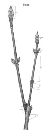
A. External Form of a
Woody Twig
Examine the woody dicot
twig provided. Note at the tip of the twig the terminal bud protected by bud
scales. Locate a node (leaf
attachment region of stem), and examine the leaf
scar(s) present. How many bundle
scars are present in each leaf scar? (Bundle scars usually appear as tiny
bumps where strands of xylem and phloem broke when the leaf fell from the stem;
if necessary, use your dissecting microscope to locate them.) Groups of small,
narrow scars (bud scale scars) often
extend around the twig; these sometimes indistinct scars are created when the
terminal bud scales of a previous terminal bud fall off. Are any such groups of
scars present? If so, how old is your twig?
Note
the small, slightly raised lenticels, located
mostly in the outer bark of internodes (regions of stem between nodes).
Lenticels consist of spongy, slightly larger cork cells that may or may not
have waxy suberin; they function in permitting exchange of gases (e.g., oxygen,
carbon dioxide) between the interior of the stem and the air.
Leaves
are usually attached to the stem at an angle. The region immediately above the
leaf base and the stem is called an axil.
Are the axillary buds (there is
normally one axillary bud in each axil) similar to the terminal buds? If not,
how do they differ?
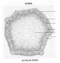
B. Herbaceous Dicot
Stems
Select a slide showing a cross section of a young alfalfa stem
(Medicago, young xs), and examine it
under low power. Identify the tissues that have been produced by the apical meristem.
These tissues include the epidermis, which
consists of a single layer of cells that covers all of the exterior of plant
organs; the cortex, consisting of
thin-walled parenchyma cells that function in food storage; primary xylem, whose vessels and tracheids conduct water and minerals; primary phloem, whose sieve-tube
members and associated companion
cells function in conduction of food in solution; and pith, whose parenchyma cells, like those of the cortex, function in
food storage. How does the arrangement of the primary xylem and phloem in this
stem differ from their arrangement in the buttercup root you examined in the
previous exercise?
Examine
an older section of alfalfa stem (Medicago,
old xs). Note that the xylem and phloem are a little more extensive, due to
the addition of secondary xylem and secondary phloem by the vascular cambium, a narrow layer of
brick-shaped, meristematic cells that develops between the primary xylem and
phloem.
With
the aid of a sharp razor blade, cut paper-thin
cross sections of a Begonia, Coleus, or
other provided stem. Mount in a drop of phoroglucinol (or water) on a slide,
and add a coverslip. Examine with the lowest power of your microscope. What
similarities between this and alfalfa stems can you detect?
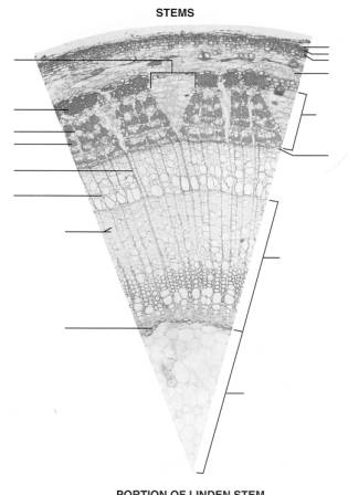
C. Internal Structure of a Woody Stem
Your instructor may have
you make your own microscope slide of a woody stem, OR he or she may choose to
have you examine previously prepared slides exclusively. If you are to make
your own preparation, one procedure is as follows:
Using
a sharp razor blade, cut paper-thin cross
sections of a 2- or 3-year-old basswood or linden (Tilia) twig. (Caution: The
wood is hard; try to brace the twig before cutting so that the razor blade
doesn't slip and injure you. You may need to practice a little to get the
sections thin enough.) Float the sections in water. Transfer the two thinnest
sections, without allowing them to become dry, to a drop of water on a slide.
Have gentian violet stain ready, and
blot off the water with filter paper. Immediately add a drop of the stain to
each section. After about 30 seconds, blot off the gentian violet stain, and
add a drop of 95%
alcohol to
each section. Wait about 1 minute, blot, and add a drop of eosin stain. After 1 more minute, add alcohol again, blot off, and
add a drop of clove oil to each
section. The slide should now be ready for the addition of a coverslip and microscopic
study.
If
you have a good slide and want to preserve it permanently, you may do so by
following the clove oil with a drop or two of xylene. Then add balsam and
place the coverslip on top. After that, the slide will probably take a few
days to dry. It can then be stored indefinitely. After you have examined your
stained sections, turn to a prepared slide of the same plant (Tilia xs) and compare it with the
slide(s) you have made.
Contrast
the linden stem with the alfalfa and Begonia
or Coleus stems. Note that the
phloem in the basswood or linden stem is quite complex, and that there are
several additional tissues. Focus on the outermost part of the basswood stem
first. Note that by the time the stem is 2 or 3 years old, the epidermis has been lost, and the cells
that are sloughing off to the outside are cork
cells produced by a cork cambium that
has developed toward the outer part of the original cortex. The cork cambium, consisting of a narrow band of
meristematic cells, also produces phelloderm
cells toward the inside of the stem. The
phelloderm cells resemble the cells of the cortex.
To
the interior of the cortex is a cylinder of phloem,
which, as previously mentioned, is quite complex in basswood or linden
stems. In cross section, it appears as a circular band of wedges alternating
with tapering trapezoids of banded tissue. The wedges include relatively large
parenchyma cells that are the flared-out tops of broad rays. The rays function in lateral conduction of water, food,
and other materials throughout the stem. The part of the ray in the phloem is
referred to as a
phloem ray, while
the part of the same ray in the xylem is called a xylem
ray. In cross section, basswood or linden stems reveal
that the broad rays are usually two or three cells wide in the xylem but flare
out and become many cells wide in the phloem. Narrow rays, however, are usually one cell wide in both the xylem
and phloem. The banded, tapering trapezoids consist of thin-walled sieve-tube members and companion cells (usually stained green)
between bands of thick-walled fiber cells
that give strength to the stem. The fibers (usually stained red or purple) are
often slightly larger at the outside edge of each trapezoid; the larger fibers
were produced by the apical meristem as part of the primary phloem, while most of the phloem visible in this slide is secondary phloem produced by the vascular cambium, a narrow band of brickshaped
cells at the base of the wedges and trapezoids.
The
vascular cambium also produces secondary
xylem toward the interior. Note that the xylem (wood) appears to have been produced in bands or rings. In fact,
each year's production of xylem by the vascular cambium is referred to as an annual ring. In basswood or linden
stems, each annual ring consists of larger vessels
that are produced when the cambium first becomes active in the spring, and
then progressively smaller vessels and tracheids produced throughout the
remainder of the growing season. Note, again, that the broad phloem rays become
broad xylem rays once they cross the vascular cambium, and that there are
several narrow phloem rays (becoming narrow xylem rays in the xylem), usually
one cell wide, between the broad rays. The large, thin-walled parenchyma cells
in the center constitute the pith.
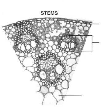
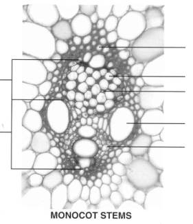
D. Monocot Stems
The alfalfa, Begonia or Coleus, and basswood stems are all dicot (dicotyledonous) stems. Examine a slide of corn (Zea mays xs) stem, representative of monocots (monocotyledonous plants).
Note that the xylem and phloem are in vascular
bundles that are scattered throughout the stem instead of being in a ring
as they are in dicot stems. Notice, also, that all the tissues are primary; there are no secondary tissues because no cambium is present to produce them. This
also results in there being no separation between cortex and pith, and the
parenchyma cells throughout which the vascular bundles are scattered are referred
to as fundamental tissue instead.
Drawings
to Be Submitted
1.
Label the provided drawing of the woody twig. The labels should include
TERMINAL BUD, AXILLARY BUD, NODE, INTERNODE, BUNDLE SCAR(S), GIRDLE,
LENTICEL(S), and BUD SCALE(S).
2.
On the illustration of the alfalfa stem provided, label the following:
EPIDERMIS, CORTEX, XYLEM, PHLOEM, PITH, and VASCULAR BUNDLE.
3.
Label the following on the drawing of a wedge-shaped portion of a cross section
of a linden stem provided: CORK, CORK CAMBIUM, PHELLODERM, CORTEX, PHLOEM,
VASCULAR CAMBIUM,
ANNUAL RING OF XYLEM,
PRIMARY XYLEM, SECONDARY XYLEM, PITH, BROAD PHLOEM RAY, BROAD XYLEM RAY, NARROW
PHLOEM RAY, NARROW XYLEM RAY, PRIMARY PHLOEM, and SECONDARY PHLOEM.
4.
Label the illustration of the monocot (corn) stem provided. Indicate VASCULAR
BUNDLE, EPIDERMIS, and FUNDAMENTAL TISSUE on the wedge-shaped portion of the
cross section. Label BUNDLE SHEATH CELL(S), SIEVE-TUBE MEMBER, COMPANION CELL,
VESSEL MEMBER, TRACHEID, XYLEM, and PHLOEM on the enlargement of a single
vascular bundle.
Questions
1. What protects the
buds of dormant twigs?
2. What are bundle scars?_______________________________________________________________
3. Where, specifically,
are axillary buds located?
4. What structures
associated with gas exchange are found throughout stem internodes?
5. What is the
difference between bud scale scars and
leaf scars?
6. Which tissue
separates cortex from pith in an older alfalfa stem? What is
the function of this tissue?
7. What is the primary
function of cortex and pith?
8. Which tissue conducts
water and minerals in solution?
9. If you saw cross sections of Begonia or Coleus and alfalfa stems side by side, what differences would be
obvious?
10. Which stains are
used to make the tissues of your handmade linden (basswood) slide more readily
visible?
11. If you wished to
make your handmade linden (basswood) slide permanent, which additional
substances would you use?
12. Which two tissues are produced by the cork cambium, and which two tissues are
produced by the vascular cambium?
1.
Where are axillary buds located?
2. What are the small
bumps of parenchyma tissue on the surface of the internodes called?
3.
How is a bundle scar formed?___________________________________________________________
4.
What is the function of a lenticel?
5. Which of the stems in
this exercise has the most complex phloem?
6. What stains are used
in making your own linden (basswood) slide?
7. In addition to cork, what tissue is usually produced by
the cork cambium?
8.
How are vascular bundles arranged in
a monocot stem?
9.
Which of the stems featured in this laboratory exercise is (are) NOT (a) dicot(s)?
10. To make your own microscope slide of a linden
(basswood) stem permanent, what substance
would you add just before placing a coverslip on it?