Roots
Materials
1.
Radish or grass seedlings germinated on damp filter paper in petri dishes
2. Prepared slides of
cross sections of young buttercup
(Ranunculus) and greenbrier (Smilax) roots
3. Prepared slides of willow (Salix) roots showing lateral
roots
Some
Sugggested Learning Goals
1.
Understand the differences between root hairs and lateral roots.
2.
Know the locations and functions of root tissues such as epidermis, cortex, endodermis, pericycle, phloem, and xylem.
3.
Know the location and composition of Casparian
strips.
Introduction
Roots function primarily
in anchoring plants and in absorbing water and dissolved substances vital to
growth and maintenance of living tissues. The typical regions of a young root
tip (root cap, meristematic region, region
of elongation) were briefly examined in Exercise 3. In this exercise we
want to concentrate on the region behind the region of elongation-the region of maturation (also referred to
as the region of differentiation or root hair zone). This region is where
the cells originally produced in the meristematic region become differentiated
into several different types, each with a specific function.
Flowering
plants, primarily on the basis of differences in flower parts, are grouped into
two large classes commonly referred to as dicots
and monocots. However, dicots and
monocots also differ in the structure of their roots, stems, and leaves. In
this exercise, we will briefly examine both dicot and monocot roots.
A. Root Hairs
Root
hairs develop in a zone a short distance behind the root cap. As new root hairs
are produced near the root cap, the older root hairs farther back die. Root
hairs greatly increase the absorptive surface of a root.
Mount about half a
centimeter of a living radish or grass seedling root, including the tip, in a
drop of water on a slide, add a coverslip, and examine under low power. (Be
sure your root tip is intact; if the tip has already been removed, discard the
seedling and take another one.) Note the numerous root hairs of various sizes. Each root hair is a part of the
epidermal cell from which it protrudes; it is not a
separate cell itself. Can you distinguish the root cap that functions primarily in
protection of the delicate membranes behind it as the root pushes through the
soil?
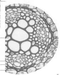
B. Dicot Roots
Examine a slide showing
a cross section of a buttercup root (Ranunculus
xs). Note that the outermost layer of cells, the epidermis, is only one cell thick. Are there any root hairs present on your slide? Next
note the extensive tissue with numerous starch
grains (often stained purple) interior to the epidermis. This tissue
functions primarily in food storage, and is known as the cortex. In carrots and similar roots, it comprises the bulk of the
root.
The
distinctive tissues in the center of the root are surrounded by a single layer
of conspicuous cells, most of which appear to have relatively thick walls. This
layer, the endodermis, forms the
inner boundary of the cortex, and separates the tissues in the center of the
root (known collectively as the stele) from
the other root tissues. The endodermis was believed to play a role in
regulating the movement of water and dissolved substances entering or leaving
the stele, but this is now in question. Endodermal cells have bands of fatty suberin (Casparian strips) around the
inner faces of the walls. Casparian strips are generally difficult to discern
because the fatty substances are dissolved when the slides are being prepared,
but in buttercup roots a conspicuous wall layer (that usually stains red) is
deposited inside the Casparian strips. Although suberin itself is impervious to
water, endodermal cell walls have many paired pits (thin areas where there is no suberin) that
allow water to pass through.
The
tissue in the center with relatively thick-walled cells (usually stained red)
is primary xylem, which functions in
conducting water. Between the arms of the xylem are patches of primary phloem, a food-conducting
tissue. In older dicot roots, a
vascular cambium usually
develops between the primary xylem and phloem, and produces secondary xylem and phloem. The addition of secondary tissues
by the vascular cambium will increase the girth of the root. Note the pericycle, a single layer of thin-walled
cells located adjacent to and inside the endodermis. The cells of the pericycle
usually do not appear different in form from a
the young primary phloem cells. Lateral
roots originate in the pericycle. Unlike dicot stems,
dicot roots have no pith.
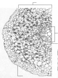
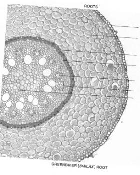
C. Monocot Roots
Examine a slide showing
a cross section of a root of greenbrier (Smilax xs), a monocot. Note the pith in the center, and
the phloem within
the patches of xylem.
Locate the endodermis,
pericycle, cortex, and epidermis. What differences and similarities are
there between dicot and monocot roots?
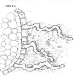
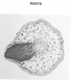
D. Lateral Roots
Examine a slide of a
cross section of a root of willow (Salix,
branching xs). This slide shows at least one specially
stained lateral root beginning to grow out from the stele. In which specific
tissue is the base of the branch root located? Which tissues does it push
through as it grows?
Drawings
to Be Submitted
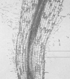
I
. Label the provided illustration of a young root through the region of
maturation. Indicate the CORTEX, EPIDERMIS, and ROOT HAIRS. Label an EPIDERMAL
CELL and a ROOT HAIR on the longitudinal section through a young root, and add
a ROOT CAP to the bottom.
2.
On the illustration of the cross section of a buttercup root, label EPIDERMIS,
CORTEX, ENDODERMIS, and STELE. On the illustration of the enlargement of the
stele, label PRIMARY XYLEM, PRIMARY PHLOEM, PERICYCLE, and ENDODERMIS.
3.
Label the following on the illustration of the cross section of a greenbrier
(SMILAX) root: EPIDERMIS, CORTEX, ENDODERMIS, PERICYCLE, PRIMARY PHLOEM,
PRIMARY XYLEM, and PITH.
4.
Label a cross section of a willow root, showing a developing lateral root.
Label EPIDERMIS, CORTEX, ENDODERMIS, PERICYCLE and LATERAL ROOT.
Review
Questions
1.
With which specific region of roots
is this exercise concerned?
2.
In which tissues do the following originate?
Root hairs____________________________
Lateral roots_______________________________________________
3. What evidence of the
food-storage function of cortex is
present in buttercup roots?________________________________
4.
Which tissue surrounds and borders the stele of a dicot root?
Which
tissues comprise the stele?
5.
What is the function of the vascular
cambium?
6.
Of what substance are Casparian strips composed?
7.
Is a pith present in all roots?______________________________________________________________________
If not, in which roots is it present?___________________________________________________________________
8. As lateral roots
develop inside a primary root, through which tissues must they grow to reach
the surface?________________
Laboratory
Preparation
Quiz 4
Roots
1. From which tissue do lateral roots arise?
2. Between which tissues is the vascular cambium located?
3.
Which tissue of stems is not present in dicot roots?
4.
In which tissues are root
hairs to be found?________________________________________________
5. Which tissue is
immediately adjacent to the endodermis
on the side toward the center?
6. In which region of
the root does differentiation of cells into various cell types take place?
7. What is present in
cells of the cortex
that gives evidence of its function as a food-storage tissue?_______
8.
Of what fatty substance are Casparian
strips composed?
9. What tissue produces
cells that add to the girth (diameter) of the root?
10. What
water-conducting tissue is present in the center of a dicot root?