Bryophytes
and Ferns
Materials
1.
Live mosses with sporophytes attached
2.
Prepared slide of moss protonema
3. Live Marchantia with archegoniophores and
antheridiophores
4. Petri dish with live
protonemata (demonstration)
5. Prepared slides of
longitudinal sections of archegonial heads of Mnium (or similar moss)
6. Live hornworts
(demonstration)
7. Variety of live fern
plants, one with expendable fronds that have mature sori
8. Live prothalli
(demonstration)
9. Prepared slides
(whole mounts) of bisexual prothalli
Some Suggested Learning Goals
1.
Understand how the development of gametangia
(structures in which sex cells are produced) and zygotes of members of the Plant Kingdom differs from the
development of gametangia and zygotes in members of other kingdoms.
2. Understand how the form and structure of bryophytes differs from that of more
complex plants.
3.
Know what develops or takes place in each phase of the life cycle of a moss.
4.
Know what develops or takes place in each phase of the life cycle of a liverwort.
5. Learn how asexual and sexual reproduction of
both thalloid and "leafy" liverworts differs
from that of mosses.
6. Be able to explain
basic differences between the sporophytes of ferns and mosses.
7. Know the life cycle
of a typical fern.
8. Understand the nature
of a prothallus and a sorus, and the roles they play in a
fern life cycle.
Introduction
In algae, fungi, and
other relatively primitive organisms that in the past were regarded as plants,
the gametes (sex cells) are produced
in single-celled gametangia, and the zygote often undergoes meiosis directly.
Beginning with the bryophytes (e.g., mosses),
however, the gametes are produced in gametangia that are composed of many
cells; the zygote, through mitosis, develops into an embryo that, in turn, develops into a diploid sporophyte. Spores are produced by meiosis within a specialized part of the sporophyte.
Alternation of
Generations in bryophytes and ferns is marked by the
development of distinct, separate gametophyte and sporophyte bodies. In
bryophytes, the sporophyte, while a distinct body in itself, is dependent on
the gametophyte for most of its nutrition. In ferns, however, both the
gametophyte and the sporophyte are photosynthetic and independent of each
other.
A.
Bryophytes (Phyla Bryophyta, Hepaticophyta, and Anthocerophyta)
Mosses, liverworts, and
hornworts are included in these three
phyla. Bryophytes differ from higher plants in Jacking xylem and phloem,
although some do have specialized cells that can conduct a little water and
food in solution. Some species may form extensive low mats consisting of dozens
or even hundreds of plants. Because true xylem and phloem are lacking, however,
none of the individual plants become very large. They cannot grow or function
very long without external moisture; hence their usual association with damp
habitats.
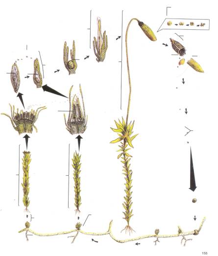
Examine
the clump of moss provided. The clump consists of small green
"leafy" gametophyte plants.
("Leafy" is in quotation marks because unlike true leaves, which are
diploid (2n), those of mosses consist of a single layer of haploid (n) cells; moss and liverwort
"leaves" also have no internal structure or stomata.) The
"leaves" do, however, carry on photosynthesis like the true leaves
of more complex plants.
Some
of the moss plants may have a thin stalk or seta
emerging from the tip. A capsule
(sporangium) develops at the free end of each seta. The seta and capsule
are diploid (2n) and constitute the sporophyte. Sporocytes (not visible
here) within each sporangium undergo meiosis,
producing spores. The sporangium
is usually partially to completely covered with a "pixie cap" called
a calyptra. The calyptra develops
from archegonial tissues and is therefore n
(haploid). When the calyptra is removed, a second, smaller (2n) cap may be seen covering the free
end of the capsule. This smaller cap, the operculum,
develops with the sporophyte and eventually pops off, allowing the spores
to disperse. Release of the spores is partially controlled by tiny peristome teeth at the rim of the
capsule; the peristome teeth, which resemble tiny, cross-ribbed shark's teeth,
move in response to changes in humidity.
Turn now to a prepared
slide labeled "Moss protonema." A
protonema is an algalike body that develops when a moss spore germinates.
Notice the chloroplasts present in each cell, and that the transverse walls of
the cells usually are not strictly at right angles to the other walls. Note,
also, the "buds" that are
developing along some of the threads. These buds become new "leafy"
gametophyte plants. Some may already have rootlike rhizoids at their bases. Rhizoids are only one cell thick; they may
anchor bryophyte plants in the same way true roots do, but like the remainder
of a moss gametophyte, they have no xylem or phloem and can absorb water slowly
only in very limited amounts. The word rhizoid
should not be confused with rhizome, which
is a term applied to the horizontal stems of ferns and higher plants.
Next
turn to a slide of moss archegonia. Archegonia
are female reproductive structures of mosses produced at the tips of female
gametophyte plants. Occasionally both male and female reproductive structures
are produced on the same plant. Each archegonium loosely resembles a tiny vase
with a narrow neck, the enlarged base itself being elevated on a short,
relatively wide stalk. Although there are usually several to many archegonia
produced at the tip of each plant, the archegonia are not always strictly
upright, and they are interspersed among sterile, multicellular hairs, called paraphyses. When microscope slides of
moss archegonia are made, very thin longitudinal sections are cut and stained.
Parts of the archegonia and paraphyses are often sliced off; because of this
you may not have a complete archegonium on your slide. You should, however, be
able to see at least one archegonium with its base intact. The cavity within
the archegonium base contains an egg.
Turn
now to a slide of moss antheridia. These
structures, before they are sliced, are shaped like miniature clubs; they
contain numerous sperms. Paraphyses
usually are present among the antheridia. In nature, a sperm swims down the
neck of an archegonium and unites with the egg, forming a zygote. As the zygote divides, it forms an embryo, which is dependent on the gametophyte for its nutrition.
The embryo then develops into a
sporophyte, consisting of a seta and capsule. Even the mature sporophyte
is still largely dependent on the gametophyte for its energy.
Liverworts have
nearly all of the reproductive structures found in mosses. Although there are
many species of "leafy" liverworts, some of the most common and bestknown
forms are thalloid. Thalloid
liverworts have flattened bodies that look a little like bright-green foliose
lichens. Examine the thalloid liverworts provided. Some, such as Marchantia, have their archegonia and
antheridia elevated above the thallus on
umbrellalike archegoniophores and
disc-shaped antheridiophores. Many
thalloid liverworts also reproduce asexually by means of gemmae, which are tiny lens-shaped pieces of vegetation produced
within gemmae cups. The gemmae cups
are scattered over the surface of the thallus. Each gemma is potentially capable
of developing into a new thallus.
Examine the
demonstration of hornworts provided.
How do the sporophytes of hornworts differ in appearance
from those of liverworts
and mosses? Are there any other apparent features that distinguish hornworts
from other bryophytes?
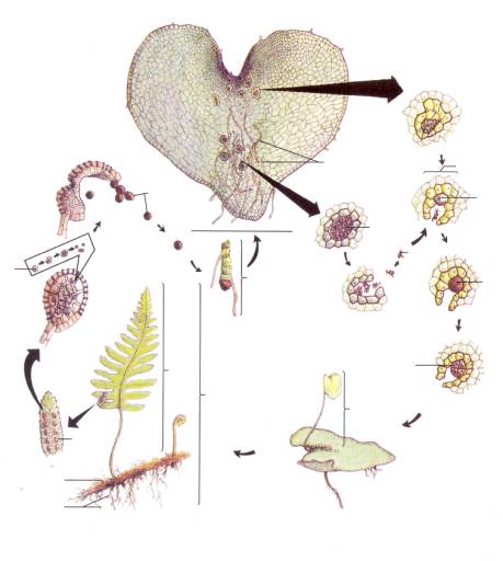
B. Ferns (Phylum Pterophyta)
Unlike the bryophytes,
the ferns do possess true conducting tissues (xylem and phloem), and the
sporophyte is the more conspicuous phase of the life cycle.
Examine
the fern plants on display. The leaves or fronds
arise from a horizontal stem (rhizome).
Notice the small brownish patches on the backs of mature fronds. Remove a
small part of a frond that has these patches and examine them with the aid of
your dissecting microscope. Each discrete patch is called a sorus and consists of a cluster of sporangia. The sporangia are often partially or wholly covered by a
transparent, umbrellalike indusium.
Sporocytes within the sporangia undergo meiosis, producing spores. The spores are released through
the springlike action of the annulus, which
is composed of heavy-walled cells around most of the edge of the sporangium.
Now
examine the green heart-shaped prothalli that
constitute the gametophytes of ferns.
Both living and preserved prothalli may be provided. Some prothalli produce
only archegonia, others only antheridia. The prothallus on the
microscope slide produces both. Find an antheridium, often located among the
rootlike rhizoids. It is circular in
outline and contains sperms. Then
find an archegonium, which is roughly the same size as an antheridium but has a
short neck. Archegonia each contain a single egg; they often tend to be close to the notch of the prothallus at
the top. In nature, a sperm unites with the egg in an archegonium, and the zygote develops into a new sporophyte, with which our study of
ferns began.
Drawings to Be Submitted
1.
Label the following on the drawings of the moss life cycle provided: MALE
GAMETOPHYTE, FEMALE GAMETOPHYTE, ANTHERIDIUM, ARCHEGONIUM, SPERM, EGG, ZYGOTE,
EMBRYO, DEVELOPING SPOROPHYTE, MATURE SPOROPHYTE, CALYPTRA, CAPSULE, SPOROCYTE,
OPERCULUM, PERISTOME, SPORES, PROTONEMA, BUD, and RHIZOIDS. Also indicate where
MEIOSIS occurs.
2.
Draw portions of sections through the tips of moss gametophytes from prepared
microscope slides, using the low power of your compound microscope. Show at
least one good ARCHEGONIUM and several PARAPHYSES in the FEMALE GAMETOPHYTE,
and at least one or two ANTHERIDIA and several PARAPHYSES in the MALE
GAMETOPHYTE. Also label EGG and SPERM(S). Be sure to reread the comments in
section A about how parts of structures may be cut off during the manufacture
of the slides.
3. Draw HABIT SKETCHES (i.e., how the organisms
appear in nature) of a thalloid liverwort and a hornwort.
4. Draw a fern SORUS, with the aid of the highest
power of your dissecting microscope. Label SPORANGIA
(and
INDUSIUM, if present).
5. Label the following on the drawings of the fern
life cycle provided: SPOROPHYTE, FROND, RHIZOME,
ROOTS, SORUS, SPORANGIUM, SPOROCYTES, SPORES,
DEVELOPING GAMETOPHYTE, (IMMATURE PROTHALLUS), PROTHALLUS, RHIZOIDS,
ANTHERIDIUM, ARCHEGONIUM, EGG, SPERM, ZYGOTE, EMBRYO, DEVELOPING
SPOROPHYTE, and YOUNG SPOROPHYTE. Also indicate
where MEIOSIS occurs.
6. Draw a fern prothallus from the prepared slide
provided. Label ARCHEGONIUM, ANTHERIDIUM, and
RHIZOIDS. (Use the lowest power of your
compound microscope.)
7. Draw a fern frond, showing the position of the
SORI.
The Lower Vascular Plant Divisions
The Fern Allies
Introduction
The vascular plants are divided artificially into
two major groups, the seedless (or spore-dispersing) vascular plants and the
seed plants. There are four major divisions of seedless vascular plants:
Psilophyta, Lycophyta, Sphenophyta, and Pterophyta. The first three divisions,
often referred to as the "fern allies" , have few living
representatives although they are well represented in the fossil record. All of
the vascular plants have a dominant sporophyte generation, and a reduced,
often, dependent gametophyte stage.
Exercise A
Psilophyta: The Whisk Ferns
The Psilophyta are represented by two living
genera, Psilotum and Tmesipteris, both of which have very simple sporophytes.
Examine the living specimens and herbarium
specimens of Psilotum . Psilotum is unique among living vascular plants because it
lacks both vascular roots and leaves. Only the stem is vascular. The scalelike
structures along the stem are called enations.
Note the dichotomous (forking) branching pattern of
the aerial portion of the plant body. The below-ground portion of the plant
axis is a rhizome (an underground stem) bearing rhizoids for the absorption of
water.
Note the three-parted sporangia, which are borne on
short side branches. Psilotum is homosporous; that is, it produces only one type
of spore. Although Psilotum is homosporous, the gametophytes are bisexual; both
archegonia and antheridia are produced on the same gametophyte. The non-photosynthetic
gametophytes develop in association with mycorrhizae.
Tmesipteris is an epiphyte that grows on tree ferns and other plants.
Lower Vascular Plants Lab - 2
Exercise B
Lycophyta: The Lycophytes
The living representatives of the Lycophyta are all
relatively small plants, with true roots, true stems, and true leaves. The
leaves are microphylls, having just one vascular connection or vein. The fossil
members of this division, however, include many woody, treelike forms (the
Lepidodendrids), which numbered among the dominant plants of the coalforming
forests of the Carboniferous period.
Examine the living specimens and herbarium
specimens of Lycopodium and Selaginella. Identify the roots, stems, and leaves (microphylls)
of these genera. Selaginella species are common in both temperate and tropical
rain forests, although it is frequently confused with mosses. Some species of Selaginella, including the "Resurrection plant", are found in very dry
habitats. Lycopodium species grow in many wooded areas throughout
temperate ecosystems.
Examine the preserved, or herbarium specimens of Isoetes. Although the leaves of Isoetes are much larger than those of Lycopodium and Selaginella, they are still microphylls. In Isoetes, the leaves are attached to a cormlike structure (a fleshy stem). Isoetes is aquatic.
The sporangia of the Lycophyta are borne on leaves
which are very similar to the sterile (non sporangia-bearing) leaves of the
plant. The sporangium-bearing leaf is called a sporophyll. In Selaginella and in most species of Lycopodium, the sporophylls occur in compact aggregates
called cones, or strobili. Examine the strobili on the specimens provided. In Isoetes , sporangia arise at the bases of the leaves, with a single sporangium
per leaf.
Lycopodium is homosporous, producing one type of sporangium. Observe the prepared
slide of Lycopodium strobilus and locate the sporangia. The
gametophytes produce both archegonia and antheridia.
Selaginella and Isoetes are heterosporous. They produce two types of
sporangia: megasporangia (female) and microsporangia (male). Spores develop
into either female gametophytes (which produce archegonia) or male gametophytes
(which produce antheridia). Observe the prepared slide of the Selaginella strobilus. Locate the larger megasporangia and the smaller
microsporangia. Compare the strobilus of Selaginella with the strobilus of Lycopodium.
It is exceedingly rare to find gametophytes of the
Lycophyta. Most are subterranean and very tiny. However, sporophyte plants
develop from the gametophyte structure so that whenever you find a sporophyte
plant, you can be assured that a gametophyte was once there. (Once a sporophyte
plant becomes established, the gametophyte degenerates.)
Lower Vascular Plants Lab - 3
Exercise C
Sphenophyta: The Horsetails
Although once a very abundant and diverse group of
plants, the Sphenophyta today are represented by a single herbaceous genus, Equisetum.
Examine the living and herbarium specimens of Equisetum. The sporophyte of
Equisetum differs from that of the other fern allies in having jointed and ribbed
stems with the leaves (microphylls) arranged in whorls at nodes.
Feel the coarse texture of the stems. The stems of Equisetum contain silica. Note that the stems, rather than the leaves of Equisetum are photosynthetic.
Examine the cones or strobili on the specimens
provided. The sporangia are borne on umbrella-like structures called
sporangiophores, rather than on sporophylls.
Examine the prepared slide of the Equisetum strobilus. Is Equisetum homosporous or heterosporous?
Examine the prepared
slide of Equisetum spores. Note that four threadlike structures,
called elaters, surround each spore. The elaters, which are sensitive to
humidity changes, are used to disperse the spores. When the sporangium breaks
open, the sudden change in humidity causes the elaters to uncoil which
"whips" the spore out of the sporangium to be carried by air current
to a new location. The gametophytes of Equisetum are green, freeliving,
and bisexual.
Questions
1.
How do moss "leaves" differ from the leaves of more complex plants?
2.
What is the difference between a calyptra and an operculum?
3. How is the release of
spores controlled in mosses?
4.
Where does meiosis take place in
mosses?
5.
Where, in mosses, are zygotes and embryos formed?______________________________________________
6. In Marchantia, what is the function of archegoniophores and antheridiophores?___________________________
7.
What are all the parts of a complete sorus?
8.
Where, specifically, are fern antheridia located?_______________________________________________
9.
What parts of a fern are 2n?_____________________________________________________________
Where,
in a fern, does the switch from 2n to
n take place?_________________________________________
Where,
in a fern, does the switch from n to 2n take place?_________________________________________
10. In addition to
seeds, what do higher plants have that bryophytes
lack?________________________________
11. Which phase in the
life cycle of a moss consists of a "leafy" plant?
12.
In which specific structure of a moss are sperms
produced?
13. What is the toothed
structure in a moss sporophyte that
controls the release of spores from a
sporangium?
14. How does a thalloid liverwort differ in
appearance from a moss?
15.
What is a cluster of fern sporangia called?___________________________________________________
16.
Where does meiosis take place in
ferns?_____________________________________________________
17.
What name is applied to the gametophyte of
ferns?______________________________________________
18. Where are fern antheridia produced (i.e., among what
structures on the gametophyte)?_____________________
19. What are the differences among rhizoids, roots, and rhizomes?