Kingdoms Archaea, Bacteria, and Protista
Materials
1.
Plates of gram-positive and gram-negative bacteria 2. Bacterial
plates showing a variety of colonies 3. Bunsen burners
4. Live and/or preserved
Anabaena or Nostoc colonies 5. Live cultures of Ulothrix, Spirogyra, Oedogonium,
Volvox, Scenedesmus,
Euglena, or Phacus
6. Slides of stained and
preserved Ulothrix, Spirogyra,
and Oedogonium
7. Diatomaceous earth
8.
Herbarium specimens of seaweeds such as Gelidium, Porphyra, Gigartina, Ulva, Codium, Postelsia,
Laminaria, Costaria, Nereocystis, or Desmarestia
9. Live or preserved dinoflagellates
10.
Loaf of sliced white bread that contains no preservatives
11. Small petri dishes
12. Pond water
13. Dropper bottles of
gentian (crystal) violet 14. Dropper bottles of safranin
0 dye 15. Dropper bottles of 95% ethyl alcohol
16. Live slime mold
plasmodia
17. Non-expendable and expendable slime mold
sporangia 18. Preserved, dried, or living specimens of slime molds
Some Sugqested
Learning Goals
1.
Know how to distinguish gram-positive bacteria
from gram-negative bacteria, and all
bacterial cells from those of Kingdom Protista.
2.
Understand distinctions between heterocysts and akinetes.
3.
Learn the differences among Ulothrix, Spirogyra, and
Oedogonium with respect to reproduction and
chloroplasts.
4.
Understand how a
diatom is
constructed and how it moves.
5.
Know the parts and structure of a dinoflagellate and
of a
kelp or other large seaweed.
6.
Know how a slime mold plasmodium moves
and the structure'of slime mold sporangia.
Note;; Slime molds (myxomycetes)
are believed to be members of Kingdom Protista
because they have several features commonly found in other members of this
kingdom, including reproductive cells with flagella, that are not generally
found in members of Kingdom Fungi. Nevertheless, because
they
also have certain funguslike features, they have in
the past been treated as fungi and have traditionally been studied along with
members of Kingdom Fungi, discussed in the next exercise (Exercise 15). Your instructor
may or may not choose to defer examining these organisms until you study
Kingdom Fungi.
Introduction
Kingdoms Archaea
and Bacteria
All the members of
Kingdoms Archaea and Bacteria have prokaryotic cells. Prokaryotic cells
have no nuclei or other organelles bounded by membranes. Bacteria occur in
three basic forms: cocci,
which are more or less spherical; bacilli, which tend to be rod-shaped; and spirilli, whose cells are twisted like corkscrews. The kingdom previously
known as Monera is divided into two kingdoms, based
on some fundamental differences in the chemistry, metabolism, and RNA
molecules of the cells. Kingdom Archaea includes
anaerobic methane bacteria, salt bacteria
that carry on a simple form of photosynthesis and live in water saturated
with salt; and sulfolobus bacteria, which live exclusively in
Nearly
all bacterial cells are considerably smaller than those of complex plants and animals,
and are best examined with the highest power of a compound microscope. We will
examine representative bacteria that have no pigments within their cells, and cyanobacteria that have chlorophyll and other pigments in
membranes within the cells.
Kingdom Protista
Members of Kingdom Protista all have eukaryotic
cells with nuclei and various organelles discussed in earlier exercises.
With the exception of protozoans, sponges, water molds, and slime molds (slime molds are the only
group of these organisms that will be discussed here), virtually all members of
Kingdom Protista possess chlorophyll and other
pigments confined to chloroplasts.
The
active state of slime
molds is called a
plasmodium. Unlike a true mold, which consists of delicate
threads that in most fungi are compartmentalized into individual cells, a
plasmodium consists of a multinucleate mass of cytoplasm without cell walls.
Plasmodia move over dead leaves and debris in a "crawling-flowing"
motion. As the plasmodia move, they engulf bacteria and other food materials.
Slime molds, like fungi, have glycogen as a primary food reserve, and other
fungus like features such as stationary reproductive bodies (sporangia). They
differ sharply from true fungi, however, in their flagellated reproductive cells-a
feature that suggests they originated from other members of Kingdom Protista.
In
this exercise you will also be introduced to a few representatives of the
thousands of species of pigmented algae.
Algae vary in size from minute single-celled organisms to
giant kelps that may attain lengths of 45 meters (nearly 150 feet). External
water is essential to algae completing their life cycles, and the great
majority of them are aquatic. All possess chlorophyll a, but each phylum exhibits
unique combinations of pigments, different food reserves, and distinctive
reproductive cells. They occur in nature as single cells, colonies, filaments, thalli
(flattened bodies), or in mutually beneficial associations
with fungi.
A. Non-Photosynthetic
Bacteria
Examine the plates of
bacterial colonies growing on agar,
a gelatin like substance obtained from several red and a few
brown seaweeds. Note the colors and textures of the colonies, which consist of
many thousands of bacteria. Mount a small
amount of bacteria in a drop of water on a slide by touching
the tip of a probe to a colony and then vigorously rotating it in the drop of
water. Cover with a coverslip and, after locating
cells under low power, switch to high power. Note the shapes and sizes of the
bacteria.
In
the nineteenth century, Christian Gram discovered that some bacteria retain a
stain he devised and others do not retain it. His stain became known as the Gram stain; those
bacteria retaining the stain were called gram-positive and those not retaining
the stain were called gram-negative.
Variations of the Gram stain are now routinely used as a first
step in identifying bacteria. If your instructor decides to have you check
bacteria provided for their response to a gram stain, he/she will show you how
to make bacterial smears.
You should then be ready to proceed as follows:
On
clean microscope
slides, make smears of bacteria in the plates marked A and B, one smear to a
slide. Dry the slides by passing them rapidly through a Bunsen burner flame four or five
times. After the slides are dry, add a drop of gentian (crystal) violet dye to
each slide. Tilt the slides `so that any excess dye
drains away from the bacteria, and add a drop of Gram's iodine reagent. Allow
the iodine to stand for 1 to 2 minutes and then add, one at a time, drops of
95% ethyl alcohol until the violet color is no longer apparent to the naked
eye. Now add one drop of safranin 0 dye and wait for
30 seconds. Then wash gently
with water,
add
a coverslip, and examine with the microscope. Note that the bacteria on one slide are stained purple and those on
the other are not. The gram-positive
bacteria are stained purple; the gram-negative are not stained.
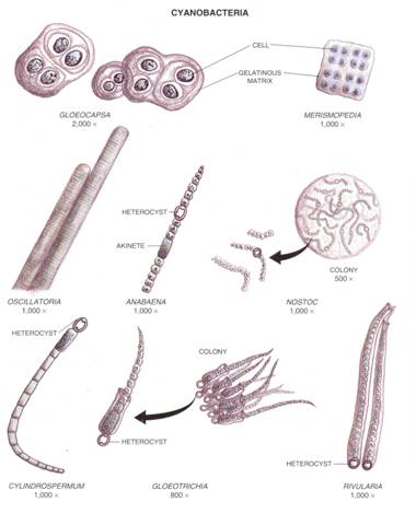
B. Cyanobacteria
Mount a small amount of the cyanobacteria Nostoc or Anabaena on
a slide or, if fresh material is not available, examine the prepared slides of
these cyanobacteria. Note that the pigments are
diffused throughout the cells and not located in plastids. Can you see any
nuclei? Are there any colorless heteorocysts (cells that appear to have a slightly
thicker wall) scattered throughout the filaments? Heterocysts
are nitrogen-fixing cells at which cyanobacterial
filaments may fragment (break). Are there any dense-looking cells that are
somewhat oblong in outline (akinetes) present at the ends or
within the filaments? Akinetes are resistant to
freezing and desiccation, and are a means of ensuring the survival of the
organisms over winter or when water is lacking.
C. Pond Water Organisms
Agitate the pond water
and place a single drop on a
clean slide; add a coverslip.
Examine with the compound microscope. Make drawings or diagrams of at least
three different organisms. To help you identify the organisms, there are
pictures of some of the more common ones on the pages at the end of this exercise.
Your instructor may help you identify other organisms not illustrated. Do you
see any motile forms
(i.e., forms that are moving)? Movement may be by means of whiplike
tails called flagella
or by means of numerous short, moving hairs called cilia. Diatoms have
a rigid glasslike cell wall composed primarily of silica; their movement may be
brought about by means of cytoplasm extending through pores and functioning
somewhat like a Caterpillar tractor track. Both flagella and cilia are minute
in diameter and may be difficult to see without special equipment or
techniques. Notice the wide variety of chloroplast types and the small, round,
colorless pyrenoids (starch accumulation
centers found in green algae) on some of the larger chloroplasts (they may or
may not be present on your particular slide).
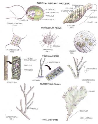
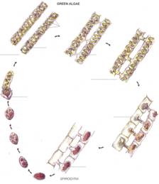
D. Spirogyra
Mount a small amount of Spirogyra in
a drop of water on a slide. Locate a pair of conjugating filaments. If your fresh material
is not conjugating, examine a prepared slide showing this. Observe the papillae that unite,
forming conjugation tubes
(short cylindrical tubes between adjacent cells). Are any gametes migrating through
the conjugation tubes to adjacent cells? If the cells of one filament are
empty, note the relatively thick zygotes
in the cells opposite those of the empty filament. No special
asexual reproductive cells are produced by Spirogyra. Instead, new cells are added by mitosis
after filaments break.
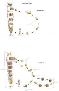
E. Ulothrix and
Oedogonium
Examine the cultures of Ulothrix and Oedogonium available, and study the prepared slides of these two green
algae. How do these two algae differ from one another and from Spirogyra with respect to chloroplasts and reproduction?
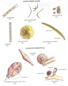
E Diatomaceous Earth
In a drop of water on a
clean slide, mount a
small amount
of diatomaceous earth, which consists
of the "shells" of millions of marine diatoms. What are the most
common shapes of marine diatoms? Is there variation in the patterns of pores
present? Note that diatoms may appear to have one shape in valve (top or bottom) view and another shape in girdle (side) view.
G. Dinoflagellates
In a drop of water on a
clean slide, mount a small amount of
the dinoflagellate material provided, or
alternatively, examine the demonstration that has been set up. Note the
"armor" plates. How are the grooves in which the
flagella are located arranged?
H. Seaweeds
Examine the herbarium
specimens of marine algae (seaweeds) on display. Although some of the larger
seaweeds have forms of food- or water-conducting tissues, none have true xylem
or phloem, root, or leaves. Do any of them have holdfasts (rootlike structures that
anchor them to rocks), bladders (bulblike
swellings that enable seaweeds to float), stipes (stalks), or blades (flattened, leaflike bodies)?
I. Slime Molds
With the aid of your
dissecting microscope, examine the petri dishes with
living plasmodia of slime molds. Focus on the leading edge
of a plasmodium and note the rapid flowing of the protoplasm. Does the
direction of flow ever change? Can you see individual cells?
Examine
the specimens of slime mold reproductive bodies available. If expendable
materials are provided, mount a
sporangium in
a drop of water on a slide. Observe the numerous spherical spores and the capillitial threads interspersed
among the spores. A few species of slime
molds lack capillitial threads, which are unknown in true fungi.
Drawings to Be Submitted
1.
Draw a group of bacterial cells from those you mounted in water. Also draw one
or two filaments of cyanobacteria such as NOSTOC or
ANABAENA. Be sure to indicate the magnifications of your drawings.
2.
Draw at least three different algae occurring in your pond water. If they are
not illustrated on the pages at the end of this exercise, ask your instructor
to identify them for you.
3.
Label the drawings of SPIROGYRA and ULOTHRIX provided. Labels for SPIROGYRA
should incude: VEGETATIVE FILAMENT, PAPILLAE,
CONJUGATING FILAMENTS, CHLOROPLAST, PYRENOID, GAMETE, ZYGOTE, and GERMINATING
ZYGOSPORE. Labels for ULOTHRIX should include: HOLDFAST, ZOOSPORES, NEW FILAMENT,
GAMETES, FERTILIZATION, ZYGOTE, MEIOSIS, and MATURE FILAMENT. Show where MEIOSIS takes place in both organisms.
4.
Draw a filament of OEDOGONIUM from a prepared slide. Label VEGETATIVE CELL,
OOGONIUM, EGG, ANTHERIDIUM, and SPERM.
5. Draw a diatom,
showing its markings.
6.
Draw a dinoflagellate showing its grooves and
"armor" plates.
7.
Draw a marine alga (seaweed). Identify any HOLDFASTS, BLADDERS, STIPES, or
BLADES present.
8.
With the aid of your dissecting microscope, draw several slime mold sporangia. Label SPORANGIUM and CAPILLITIAL THREADS.
When you have completed
your assignments for this exercise, break off a portion of bread small enough
to fit within the petri dish provided. Then add no more than one
drop of
water to the bread (if you add more than one drop, yeasts are very likely to
multiply and interfere with the growth of other fungi you will be trying to
cultivate). Next, sprinkle dust from the corners of the floor in the laboratory
or elsewhere, or comb your hair over the bread. Close the dish and print your
name and section number on the outside, then set it aside until the next
laboratory session. At that time we will examine any fungi that develop on the
bread.
1. What is the
difference between prokaryotic and eukaryotic cells?
2.
If a bacterium is gram-positive, how
does it respond to a Gram stain?
3. Which organisms are
most likely to contain heterocysts?
4. Of what advantage to
a cyanobacterium is an akinete?
5. What is the function
of conjugation tubes?
6.
Specifically where in a cell are pyrenoids located?___________________________________________
7.
What substance gives rigidity to diatom cell
walls?___________________________________________
8. How do diatoms move?
How do dinoflagellates move?
9. How does the plasmodium of slime mold differ from the
mycelium of a true fungus?__________________
10.
What is the equivalent of a root in a seaweed?
Kingdoms
1.
What is a pyrenoid?__________________________________________________________________
2.
In which alga would you expect to find conjugation
tubes?
3. If you find a pair of algal
filaments conjugated but the cells of one of them are empty, what are the dark
objects in the cells of the other filament?__________________________________________________________
4. What are the colorless, slightly thicker-walled cells
present in some cyanobacteria?__ 5. What is an akinete?__________________________________________________
6. In addition to differences in the way they
reproduce, what should help you distinguish Spirogyra
from Oedogonium?
7.
What is a motile alga?_________________________________________________________________
8. What type of organism
mentioned in this exercise may have bladders?
9.
Where are the pigments of cyanobacteria located?
10.
Which algae have rigid, glasslike walls?___________________________________________________
ALGAE LAB
AIM: To make yourself familiar with specimens of
algae. As with all other groups of
organisms, certain species of algae are found in certain habitats. The purpose of this material is to
demonstrate that you can identify some of the more common species and learn
where some of these species are commonly found.
MATERIALS: Microscopes, algae samples, herbarium samples
PROCEDURE: Examine
specimens of the different groups of algae noting the distinguishing
characteristics of each.
DIVISION CHLOROPHYTA
The Chlorophyta
are the green algae. They are an ancient
group, possibly extending back to the origin of photosynthetic, cellular
plants. Complex green plants are
considered to have arisen from green algae.
Distinguishing
Characteristics:
1.
Pigments---chlorophyll a and auxiliary pigments, chlorophyll b and carotinoids (yellow and orange pigments).
2.
Food reserve--true starch.
3.
Cell wall--cellulose..(exceptions)
4.
Flagellation--when present, always two to four; always anterior.
I. Examine the various specimens on demonstration.
Draw and Describe
one macroscopic ON your paper. Use
the following terms to help with the description;
Branching, cell shape, color, chloroplasts..(shape, size, location),
filaments, presence of any reproductive structures, any outer sheath present
and anything else you can describe it with!
DIVISION PHAEOPHYTA
Most brown algae (Phaeophyta)
grow in the intertidal zone. Nearly all are marine. There are no unicellular genera. Forms vary from simple branched filaments to giant seaweeds
over 60 meters long. Many of the giant
species, especially the kelps, show a high degree of external differentiation
into a root-like HOLDFAST, a short, stem like STIPE, and a long strap-like
BLADE. All larger species have air
bladders of various sizes.
Distinguishing
characteristics are:
1. Pigments--chlorophyll a and auxiliary
pigments, chlorophyll c, carotinoids and fucoxanthin (brown pigment).
2.
Food reserve--laminarin and mannitol.
3.
Cell wall of cellulose and algin (a
commercially valuable compound).
4.
Reproductive cells with two laterally placed flagella.
5.
Well developed alternation of generations.
II. Examine the various specimens on
demonstration. Draw and Describe one macroscopic sample on your paper. Use the following terms to help with the
description;
Branching, cell shape, color, chloroplasts..(shape, size, location), presence
of any reproductive structures, any outer sheath present and anything else you
can describe it with!
DIVISION
RHODOPHYTA
The red algae (Rhodophyta)
are relatively small plants, most species being less than 0.7 meters long. Their growth forms are simple filaments,
highly branched filaments or sheet-like bodies.
They are abundant in warm marine waters. A few are fresh water. They are capable of living at depths greater
than those of any other algae.
Distinguishing
characteristics:
1.
Pigments--chlorophyll a and the auxiliary, pigments, chlorophyll d, phycoerythrin, and phycocyanin.
2.
Food reserve--floridean starch.
3.
Cell wall of cellulose sometimes covered with gelatinous material
commercially known as AGAR.
4.
Absence of any kind of motile cells.
5.
Complex life cycles in many.
III. Examine the various specimens on
demonstration. Describe one macroscopic
and one microscopic sample on your paper.
Use the following terms to help with the description;
Branching, cell shape, color, chloroplasts..(shape, size, location), presence
of any reproductive structures, any outer sheath present and anything else you
can describe it with!
DIVISION CHRYSOPHYTA (golden-brown
algae (diatoms))
The golden-brown algae (Chrysophyta) possess evolutionary trends in size increase
some of which are exhibited by filamentous and colonial forms. We will examine diatoms, either filamentous
or unicellular forms. Diatoms are
characterized by cell walls composed of two overlapping halves that fit
together in a manner similar to the parts of a Petri dish.
ALGAE LAB
DRAWING SHEET
NAME______________________________
PD______
Label
all parts ith holdfast, stipe,
blade, name of speciman, collection point if
included.
Chlorophyta (Green Algae) Macroscopic
Phaeophyta (Brown Algae) Macroscopic
Rhodophyta (Red Algae) Macroscopic Rhodophyta (Red
Algae) Microscopic
Chrysophyta (Diatoms) Microscopic