Dissection of a Small Mammal
Note: Both safety goggles and a lab
apron should be worn
for this lab!
1. Wash off the specimen if this has
not been done to remove any additional preservative fluid.
2. Place specimen
in a dissection tray. Tie a piece of twine to a front and back leg. Pass the
twine underneath the dissection pan to the other side and tie it to the leg
on the opposite side to hold the legs apart.
3. Identify the sex of your animal. Link to diagram of male
V. female external structures in the
fetal pig.
4. Make incisions to open the
thoracic and abdominal cavities. This should be done
with scissors. Use the diagram of the ventral cuts of a fetal pig dissection to assist this.
Do not use razor blades or a scalpel!. Keep the scissors parallel to the skin surface to
prevent damage to the internal organs. Remove the flaps of skin to reveal the internal organs. While most of the pig's
skeleton is cartilage as it is a fetal pig, bone
development has started in the chest or thoracic area. This means
that more careful force will be required to cut through the sternum
(breast bone).
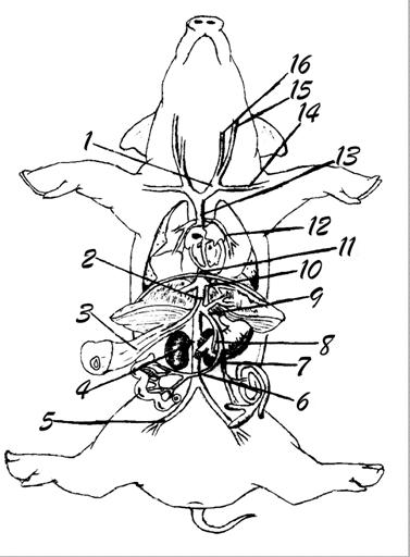
5. Identify at least 12 major
internal organs in your fetal pig. The following fetal pig diagram link may be helpful.
Link to the fetal pig heart anatomy
The period of pregnancy or gestation for
pigs is 112-115 days (3 months, 3 weeks, 3 days) and each female may produce a
litter of 7-12. As the period of development proceeds, the pig
embryos get longer, so an approximate age may be calculated from the
length. (from Odlaug: Laboratory
Anatomy of the Fetal Pig, William C. Brown, 1955.)
|
Time from Conception |
Pig Length in mm |
|
21 days |
11 mm |
|
35 days |
17 mm |
|
49 days |
28 mm |
|
56 days |
40 mm |
|
100 days |
220 mm |
|
114 days (birth) |
300 mm |
Questions
All Students
1.)
Diagram the locations of the major incisions on the ventral side of the fetal
pig.
2.) How does the position of the brain and nerve cord differ
in the fetal pig from that of the earthworm and grasshopper?
3.) What is the gestation period of the pig? How
old was your pig approximately when it was delivered from the sow? (or how would you determine an approximation of this?)
4.) How does the growth rate of the last third of a fetal
pig's development compare with that of a human embryo in the last trimester of development.
5.) State at least one function for the listing of organs
below. Indicate the system the organ belongs to (transport,
endocrine, excretory, nervous, digestive, etc.)
ex. dorsal nerve cord -- carries nerve
impulses (brain to body and vice versa) (nervous)
|
Organ |
Function |
Body System |
|||
|
brain medulla |
|
|
|||
|
brain cerebrum |
|
|
|||
|
brain cerebellum |
|
|
|||
|
heart |
|
|
|||
|
lungs |
|
|
|||
|
thymus gland |
|
|
|||
|
coronary arteries |
|
|
|||
|
diaphragm |
|
|
|||
|
stomach |
|
|
|||
|
liver |
|
|
|||
|
gall bladder |
|
|
|||
|
pyloric (stomach) sphincter valve |
|
|
|||
|
small intestine |
|
|
|||
|
esophagus |
|
|
|||
|
large intestine |
|
|
|||
|
kidneys |
|
|
|||
|
ureters |
|
|
|||
|
spleen |
|
|
|||
|
pancreas |
|
|
|||
|
testes/ovaries |
|
|
|||
|
5.
scrotum 7.
anus 8.
urogenital opening (female) 9.
urogenital
opening (male) 10.
mammary papillae 11.
tip of sternum
|
|
|
||

Next, we will go on to the respiratory system of the fetal pig.

THE MAJOR ARTERIES OF THE FETAL PIG
|
3.
right auricle 4.
renal 5.
dorsal aorta 6.
umbilical 10.
coronary 11.
pulmonary 12.
aortic arch 13.
left subclavian 14.
brachiocephalic 15.
common carotid |
|

MAJOR VEINS
|
3.
umbilical 4.
renal 5.
common iliac 6.
superior (anterior) mesenteric 8.
gastric 10.
hepatic 12.
pulmonary 14.
left subclavian 15.
external jugular 16.
internal jugular |
|
THE HEART (DORSAL VIEW)
(Please
note that some of the structures are internal and are not currently viewable,
but the arrows indicate their approximate location.)
|
2.
aorta 6.
apex 7.
right auricle 8.
left auricle 10.
left atrium 11.
bicuspid valve 12.
chordae tendinae 13.
papillary muscle 14.
tricuspid valve 15.
semilunar valve |
|

ORAL CAVITY AND PHARYNX
|
1.
hard palate 2.
soft palate 3.
nasopharynx 4.
esophagus 5.
glottis 6.
epiglottis 7.
tongue |
|

ABDOMINAL ORGANS
|
1.
gall bladder 2.
diaphragm 3.
bile duct 4.
duodenum 5.
mesentery 7.
anus 8.
rectum 9.
cecum 10.
colon 11.
pancreas 13.
stomach 14.
spleen 15.
esophagus 17.
umbilical vein |
|

THE REPRODUCTIVE SYSTEM (MALE)
In this section of our virtual disection, we will
explore the male reproductive system.
|
3.
prostate
location 6.
urogenital
opening 7.
penis 8.
urethra 9.
bulbourethral (Cowper's) gland 10.
epididymis 11.
testis 12.
vas deferens 14.
genital artery
15.
dorsal aorta 16.
ureter 17.
renal artery 18.
renal vein 19.
kidney |
|

THE REPRODUCTIVE SYSTEM (FEMALE)
In this section of our virtual disection, we will
explore the female reproductive system.
|
1.
kidney 2.
genital artery
3.
ureter 5.
cervix 7.
urethra 11.
body of uterus 12.
uterine horn 13.
oviduct 14.
ovary 15.
renal artery 16.
renal vein |
|

http://mail.fkchs.sad27.k12.me.us/fkchs/vpig/EXTERNAL ANATOMY OF THE FETAL PIG
Before
beginning to actually dissect your fetal pig, you should become very familiar
with its external anatomy. Review ANATOMICAL
TERMINOLOGY prior to starting this portion of the lab. Especially
review terms that refer to relative locations or directions
(click on Anatomical References at this website).
Procedure:
1.
Locate each of the fetal pig's anatomical
structures mentioned in the following five paragraphs.
The body is composed of
head, neck, trunk, and tail. The cut umbilical cord extends from the
ventral portion of the abdomen. The external opening of the large intestine,
the anus, is located immediately under the
tail. On the head are external ears, eyes with upper and lower lids, and a
large mouth. The tongue can be seen protruding from the mouth. The nostrils lie
dorsal to the mouth.
The first digit or toe
of both forelimbs and hindlimbs is absent and the second and fifth
digits are reduced in size. In the forelimb, the wrist (structure C) lies just above the
digits. The elbow (structure D) can be felt as a bony
protuberance on the posterior face of the leg close to the junction of the leg
with the body. The shoulder is located well above the elbow but is not
recognizable by any external feature.
In the hindlimb, the ankle (structure F) is seen as a
protuberance a short distance above the digits. The knee (structure E) is located on the
anterior face of the hindleg on about the same level as the elbow in the
foreleg. The hip can be identified by feeling a bony mass close to the middorsal
line.
Determine the sex of your specimen, and of other fetal
pig specimens in the room The male is identified by a swelling, the scrotal sac (scrotum), at the caudal end of
the body between the upper ends of the hindlegs and by the urogenital opening just caudal to the umbilical cord.
It may also be possible to feel the penis, a long muscular tubular structure
lying under the skin and proceeding caudally from the urogenital opening.
In the female, the urogenital opening is located beneath the tail
immediately ventral to the anus. A small fleshy genital papilla projects from the
urogenital aperture. In both males and females, there are two rows of mammary papillae on the ventral surface of
the abdomen.


http://www.rit.edu/~gtfsbi/genbiol/Lab%204a.htm